
Mataram University under the Indonesian Ministry of Education and Culture that provides a journal publications from research results in various fields of science from researchers in the world. The research results can be accessed freely (Open Access) so that anyone can benefit from reading and reused for research. Detailed information on journal publications see below
Journals
-
Jurnal Pepadu
JURNAL PEPADU adalah jurnal ilmiah nasional yang mempublikasikan artikel yang ditulis berdasarkan berbagai kegiatan yang dilakukan dalam rangka meningkatkan taraf kehidupan masyarakat baik di dalam negeri maupun luar negeri.
-
Journal of Fish Nutrition
The Journal of Fish Nutrition (JFN) has an objective to publish and provide high-quality scientific contributions to the field of fish nutrition. These contributions are sourced from innovative research that encourages science and technology development in the field of fish nutrition on a national and international scale. This journal serves as a communication medium for researchers, academics, students, and communities.
-
Kopula: Jurnal Bahasa, Sastra, dan Pendidikan
Journal title : KOPULA: Jurnal Bahasa, Sastra dan Pendidikan Initials : KOPULA Editor-in-chief : Dr. Burhanuddin, M.Hum. Online ISSN : 2987-5307 DOI Prefix : 10.29303/kopula Indexing : Google Scholar and view more Peer Review Process : Double-blind Frequency : 4 issues per year ( March, June, September and December) Publisher : PS. Magister Pendidikan Bahasa Indonesia FKIP Universitas Mataram Citation Analysis : Dimensions, Google Scholar Language : English & Indonesian Kopula: Jurnal Bahasa, Sastra Dan Pendidikan is a journal published 4 times a year (March, June, September, and December) published by the Master of Indonesian Language Education Study Program, FKIP, University of Mataram. Kopula was first published in 2019 as a forum for publishing the results of thoughts and research on Language, Literature, and Education. This journal applies an open access peer-reviewed journal system for both Indonesian and English articles in print and electronic versions, each of which has an ISSN. Kopula: Journal of Language, Literature, and Education has been accredited by the Ministry of Education, Culture, Research, and Technology of the Republic of Indonesia Decree No. 72/E/KPT/2024 with Sinta 5 Accreditation. Please send your articles through Online Submission. Make sure your manuscript matches the Kopula: Journal of Language, Literature, and Education template at: Volume 8 Number 2 June 2026 Edition
This journal provides immediate open access to its content on the principle that making research freely available to the public supports a greater global exchange of knowledge.
-
Journal of Fish Health
The Journal of Fish Health (JFH) has an objective to publish and provide high-quality scientific contributions to the field of fish health. These contributions are sourced from innovative research that encourages science and technology development in the field of fish health on a national and international scale. This journal serves as a communication medium for researchers, academics, students, and communities. This journal is published four times a year in February, May, August, and November since 2024.
-
Jurnal Pengabdian Perikanan Indonesia
Jurnal Pengabdian Perikanan Indonesia (JPPI) adalah jurnal ilmiah yang memuat hasil pelaksanaan kegiatan pengabdian kepada masyarakat pada bidang perikanan dan kelautan.Jurnal Pengabdian Perikanan Indonesia merupakan jurnal ilmiah yang mempublikasikan hasil kegiatan pengabdian kepada masyarakat di bidang perikanan, kelautan, dan pemberdayaan masyarakat pesisir. Jurnal ini menjadi wadah bagi akademisi dan praktisi untuk menyebarluaskan inovasi, penerapan teknologi, serta model pemberdayaan yang memberikan manfaat nyata bagi masyarakat.
-
Jurnal Ilmiah PENDAS: Primary Educational Journal
JURNAL ILMIAH PENDAS: PRIMARY EDUCATION JOURNAL merupakan jurnal yang diterbitkan oleh Program Studi Pendidikan Guru Sekolah Dasar, FKIP Universitas Mataram. Terbit dua kali dalam setahun yaitu pada bulan Juni (Januari-Juni), dan Desember (Juli-Desember). Berisi artikel artikel hasil penelitian dan atau artikel konseptual hasil kajian analitis kritis dalam lingkup Pendidikan Dasar. Fokus kajian utama Jurnal Pendas yaitu isu-isu utama dalam Belajar dan Pembelajaran, Evaluasi Pembelajaran, Metode-Media-Strategi Pembelajaran, Psikologi dan Bimbingan Konseling, Manajemen Sekolah, Bahasa Indonesia, IPA, IPS, Matematika, PPkn, dan SBdP di Sekolah dasar.
-
Darma Diksani: Jurnal Pengabdian Ilmu Pendidikan, Sosial, dan Humaniora
Darma Diksani: Jurnal Pengabdian Ilmu Pendidikan, Sosial, dan Humaniora adalah jurnal pengabdian terakreditasi nasional SINTA 4 , merupakan wahana untuk menerbitkan dan menyebarluaskan laporan hasil dari program pengabdian atau layanan kepada masyarakat, maupun penelitian berbasis pengabdian masyarakat yang berorientasi pada pengembangan teori keilmuan, konsep pemikiran, model, atau hasil penelitian yang ditujukan untuk meningkatkan kualitas hidup masyarakat. Jurnal ini menerbitkan naskah-naskah hasil pengabdian dari bidang ilmu pendidikan, sosial, humaniora, maupun bidang ilmu lain yang relevan dalam bingkai pengabdian kepada masyarakat.
-
Jurnal Rimba Lestari
Jurnal Rimba Lestari (JRL) is a periodic scientific article covering all aspects of forestry and environment sciences which includes, but not limited to the following topics: forest planning, forest policy, forest resources utilization, forest ergonomics, forest ecology, forest inventory, silviculture, and management of regional ecosystems. It is primarily a medium for disseminating original theoretical and experimental researches, as well as technical reviews. This journal issues one volume annually consists of two issues that delivered every May, and October. JRL is published by the Department of Forestry-University of Mataram.
-
-
Portal ABDIMAS
PORTAL ABDIMAS, merupakan Jurnal Pengabdian kepada Masyarakat sebagai sarana publikasi bagi para akademisi dan pihak-pihak terkait dalam bidang pengembangan dan penerapan IPTEKS yang berhubungan dengan keteknikan, lingkungan, biosains, kesehatan, gizi, agroindustri, agrobisnis, sosial, ekonomi, humaniora dan lain lain. Artikel dapat ditulis dalam Bahasa Indonesia maupun Bahasa Inggris. Tulisan harus asli, bukan merupakan terjemahan atau saduran dari artikel lain yang belum dan tidak akan dipublikasikan dalam jurnal yang lain. Jurnal PORTAL ABDIMAS diterbitkan dua kali setahun (Edisi bulan April dan Oktober).
-
Journal of Microbiology, Biotechnology and Conservation
First published online in 2025 with a focus on Microbiology, Biotechnology and Conservation published 3 (three) times a year in January, May, September.
-
Jurnal Riset Pemasaran
Jurnal RISET PEMASARAN merupakan jurnal yang dimaksudkan sebagai media terbitan kajian ilmiah hasil penelitian, pemikiran dan kajian analisis-kritis mengenai isu Ilmu - ilmu Manajemen Pemasaran dan lain-lain yang terkait. Artikel ilmiah dimaksud berupa kajian teori (theoritical review) dan kajian empiris dari ilmu terkait, yang dapat dipertanggungjawabkan serta disebarluaskan secara nasional maupun internasional.
Tujuan Jurnal Riset Pemasaran adalah:
1. Menyebarluaskan pengetahuan, penemuan, dan pengembangan ilmu pemasaran secara teori maupun praktis melalui hasil-hasil riset.
2. Mengakomodasi hasil-hasil riset ilmiah denga dunia bisnis yang membutuhkan.
3. Memotivasi para peneliti pemasaran dan praktisi bisnis untuk mempublikasikan hasil temuan dalam bidangmya. -
J-AGENT (Journal of Agricultural Engineering and Technology)
J-AGENT (Journal of Agricultural Engineering and Technology) is a scientific journal that publishes research results in the field of agricultural engineering and technology. J-AGENT's scope includes: agricultural environmental engineering and conservation, agricultural tools and machinery, bioprocess engineering, new renewable energy, agricultural information technology, smart agriculture and other related fields related to agro-industrial technology.
This journal is published by the Faculty of Food Technology and Agroindustry, University of Mataram. Articles published on J-AGENT can be written in 2 languages, namely Indonesian or English. J-AGENT (Journal of Agricultural Engineering and Technology) is published 4 times a year in January-March, April-June, July-September, and October-December (1 volume, 4 issues).
-
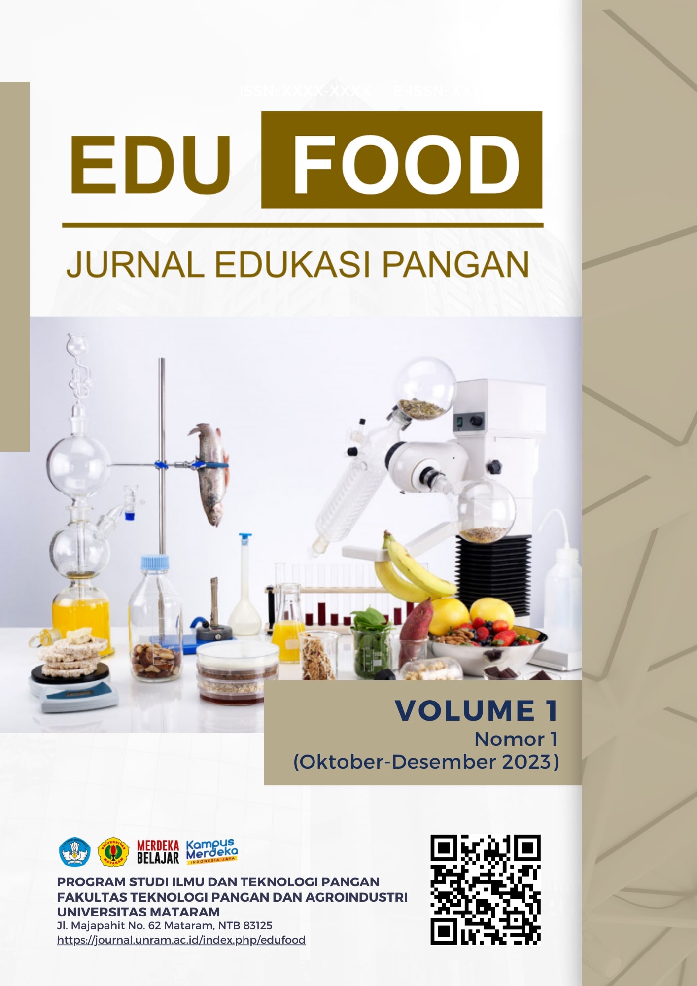
Jurnal Edukasi Pangan
Jurnal Edukasi Pangan is a peer-reviewed journal that publishes various research articles in the field of food science and technology and their applications in the food industry. This journal is managed by the Food Science and Technology Study Program (ITP) and published by the Faculty of Food Technology and Agroindustry, University of Mataram.
-
Jurnal Riset Keuangan
Tujuan Jurnal Riset Keuangan adalah:
1. Menyebarluaskan pengetahuan, penemuan, dan pengembangan ilmu Manajemen Keuangan secara teori maupun praktis melalui hasil-hasil riset.
2. Mengakomodasi hasil-hasil riset ilmiah denga dunia bisnis yang membutuhkan.
3. Memotivasi para peneliti Manajemen Keuangan dan praktisi bisnis untuk mempublikasikan hasil temuan dalam bidangmya. -
Jurnal Penelitian MSDM
Tujuan Jurnal Penelitian MSDM adalah:a
1. Menyebarluaskan pengetahuan, penemuan, dan pengembangan ilmu MSDM secara teori maupun praktis melalui hasil-hasil riset.
2. Mengakomodasi hasil-hasil riset ilmiah denga dunia bisnis yang membutuhkan.
3. Memotivasi para peneliti MSDM dan praktisi bisnis untuk mempublikasikan hasil temuan dalam bidangmya -
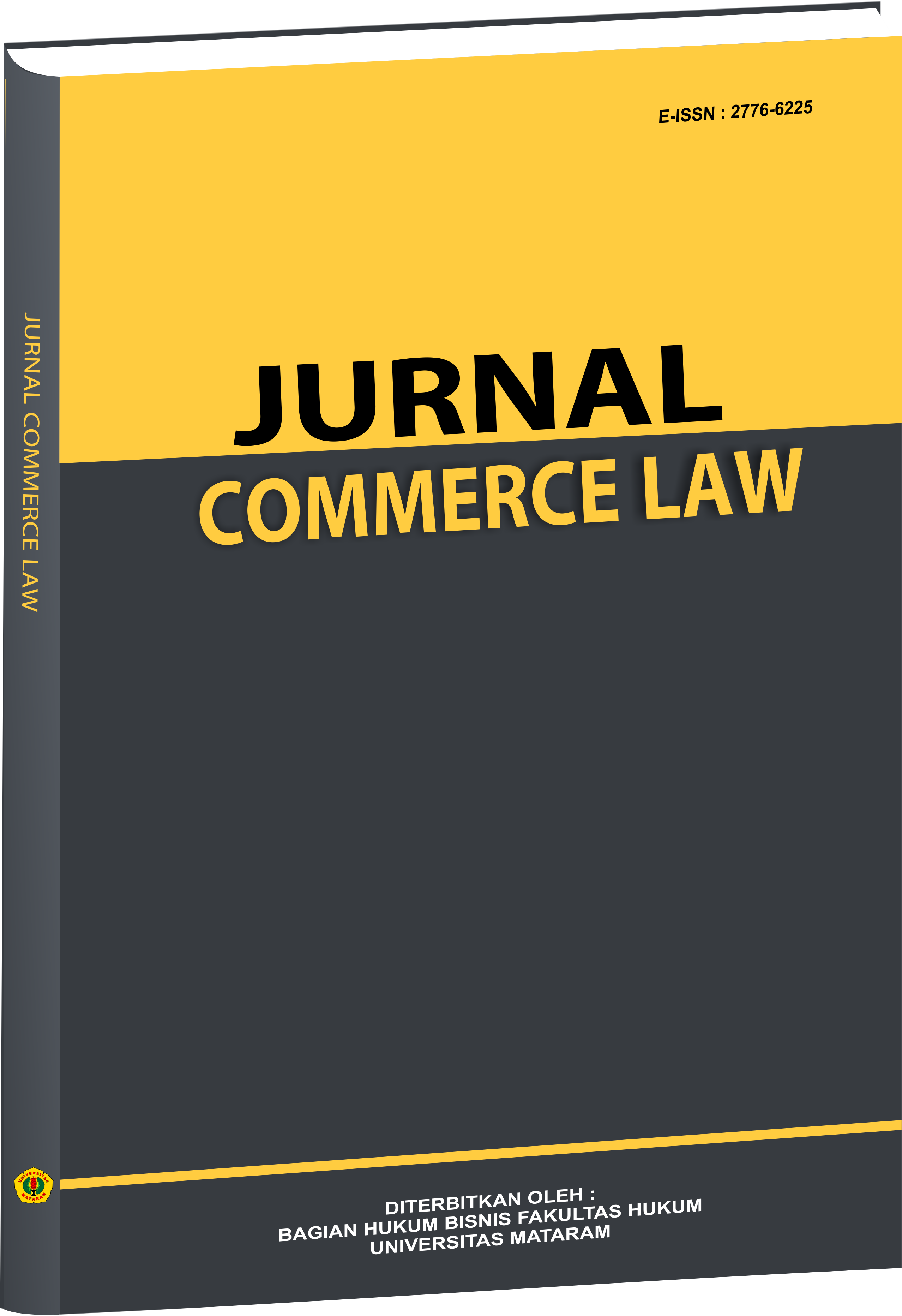
Commerce Law
Commerce Law is a medium published every semester for the dissemination of research and service results or conceptual studies on law and business matters. Commerce Law is published twice a year (June and December). Commerce Law publishes research and community service results or conceptual studies (thought results) on law, constitution, notary, and legal issues that have not been published in other media. Commerce Law is intended for experts, academics, practitioners, state officials, NGOs, and other legal observers.
-
Jurnal Diskresi
Jurnal Diskresi merupakan media open access setiap semester guna penyebarluasan (diseminasi) hasil penelitian dan pengabdian atau kajian konseptual tentang hukum. Jurnal Diskresi terbit dua nomor dalam setahun (Juni dan Desember) Jurnal Diskresi memuat hasil penelitian dan pengabdian atau kajian konseptual (hasil pemikiran) tentang hukum, konstitusi, serta isu-isu hukum tata negara yang belum pernah dipublikasikan di media lain. Jurnal Diskresi ditujukan untuk kalangan pakar, akademisi, praktisi, penyelenggara negara, LSM, serta pemerhati hukum konstitusi dan ketatanegaraan.
-
Samota Journal of Biological Sciences
SJBIOS is the official publication of the Department of Biology, Faculty of Mathematics and Natural Sciences, University of Mataram. All articles published in SJBIOS are peer-reviewed and published online for immediate access and citation.
-
Sinergi dan Harmoni Masyarakat MIPA
Jurnal SINONIM (e-ISSN 3089-736X) adalah jurnal Pengabdian kepada Masyarakat yang dikelola oleh Badan Pertimbangan Penelitian dan Pengabdian Fakultas (BP3F), Fakultas Matematika dan Ilmu Pengetahuan Alam, Universitas Mataram. Jurnal ini didedikasikan untuk hasil publikasi kegiatan Pengabdian kepada Masyarakat yang dilakukan oleh akademisi, mahasiswa, peneliti, praktisi, aktivis, dan lembaga pemerintah atau swasta dalam program CSR (Corporate Social Responsibility). SINONIM adalah jurnal akses terbuka dan semua artikel yang diterbitkan tersedia dalam versi online sebanyak 2 kali setahun (April dan Oktober)
-
Energy, Materials and Product Design
Energy, Materials, and Product Design is an open-access journal published by the Department of Mechanical and Industrial Engineering, Faculty of Engineering, University of Mataram, Mataram, West Nusa Tenggara, Indonesia. Energy, Materials, and Product Design publishes two articles per volume annually, published in May and November.
Energy, Materials, and Product Design is a forum for publishing research results, case studies, and reviews related to mechanical engineering and its intersection with industrial engineering, such as energy, materials, and product design. This includes research or case studies related to heat exchangers, energy conversion (including solar, wind, marine, and biomass), heat storage, materials and components (including manufacturing and metallurgy), electric vehicles or their components, and ergonomics applications in tool and workstation design, occupational health and safety (K3), productivity, production and manufacturing systems, work design and reliability, material handling, optimization and simulation, quality control and quality engineering, and other areas of engineering that may impact other engineering fields.
Articles may be written in Indonesian or English in accordance with the Energy, Materials, and Product Design journal guidelines.
Energy, Materials and Product Design has been indexed in the Crossref (DOI), Google Scholar, and Digital Reference Garba (GARUDA).
ISSN 2964-6987 | DOI prefix 10.29303/
-
Parhesia
PARHESIA berasal dari bahasa Yunani, merupakan istilah yang berkembang dalam ilmu hukum, yang berarti "berani menyampaikan kebenaran".
PARHESIA merupakan media jurnal elektronik ilmiah yang dikelola oleh Bagian Hukum Pidana Fakultas Hukum Universitas Mataram.
Fokus Jurnal PARHESIA pada publikasi hasil penelitian, kajian, dan gagasan pengembangan ilmu pengetahuan bidang Pidana, baik Hukum Pidana Nasional dan Hukum Pidana Internasional
-
Jurnal Pengabdian Inovasi Masyarakat Indonesia
Adalah jurnal peer-review yang diterbitkan oleh Program Studi Pendidikan Kimia, Fakultas Keguruan dan Ilmu Pendidikan, Universitas Mataram. Jurnal ini merupakan jurnal open access. Jurnal ini memuat artikel hasil pengabdian kepada Masyarakat dan penerapan inovasi, model atau konsep dan atau implementasinya dalam rangka peningkatan partisipasi masyarakat dalam pembangunan, pemberdayaan masyarakat atau pelaksanaan pengabdian kepada masyarakat. Jurnal Pengabdian Inovasi Masyarakat Indonesia (JPIMI) terbit 2 (dua) kali setahun. JPIMI sudah terakreditasi sinta 5 dengan nomor SK 177/E/KPT/2024 sejak bulan Februari tahun 2022.
-
Lombok Medical Journal
Lombok Medical Journal (LMJ) is scientific, peer-reviewed, and open access journal published by Faculty of Medicine, Universitas Mataram, West Nusa Tenggara, Indonesia.
-
The Journal of Sustainability, Economics, and Social Science
First published online in 2025 with a focus on The Journal of Sustainability, Economics, and Social Science (JSESS) published 2 (two) times a year in March and September.
-
-
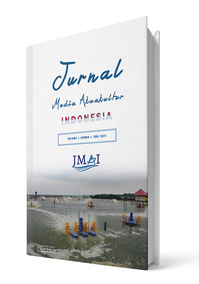
Indonesian Journal of Aquaculture Medium
Jurnal Media Akuakultur Indonesia (Indonesian Journal of Aquaculture Medium), abbreviated as JMAI (IJAM), managed by Aquaculture Study Program, University of Mataram, is a scientific journal publishing research results in marine and fisheries sector.
In 2021 (Volume 1) and 2022 (Volume 2), JMAI published twice a year.
But since 2023 (Volume 3), JMAI Published four times a year (February, May, August and November).
This development is recorded and reported on the ISSN number attached to JMAI.


-
Private Law
The Private Law Journal (ISSN 2775-9555) is a peer-reviewed academic journal published by the Faculty of Law at the University of Mataram. Established in 2021, the journal focuses on civil law and related legal studies, providing a platform for scholars, practitioners, and students to publish original research and legal analyses.
-
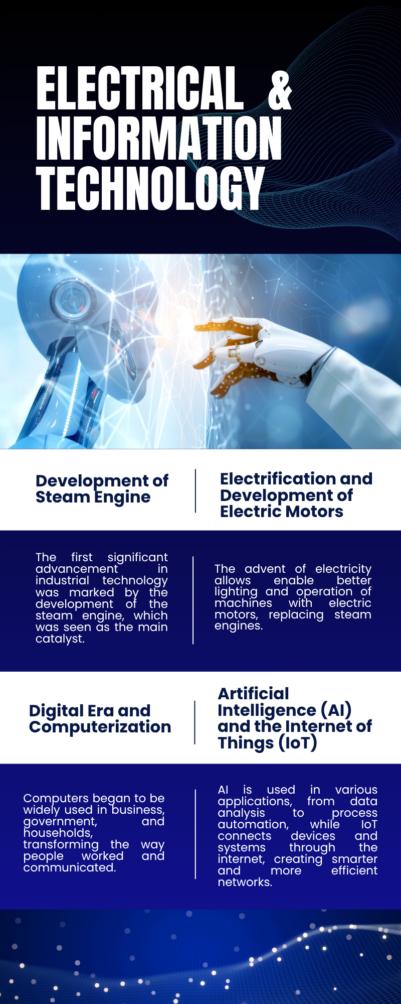
JEITECH (JOURNAL OF ELECTRICAL ENGINEERING, INFORMATION TECHNOLOGY, CONTROL ENGINEERING, AND ROBOTIC)
JEITECH : JOURNAL OF ELECTRICAL ENGINEERING, INFORMATION TECHNOLOGY, CONTROL ENGINEERING, and ROBOTIC is a journal in Electrical Engineering, Electronic Engineering, Telecommunications Engineering, Computer Engineering, Information Technology, Control and Robotic published by the Department of Electrical Engineering, University of Mataram. This journal is available to researchers who wish to improve their knowledge in a particular field, and intend to disseminate knowledge as a result of research.
-
Bio-Educ: Jurnal Pengembangan dan Inovasi Pembelajaran Biologi
Bio-educ: Jurnal Pengembangan dan Inovasi Pembelajaran Biologi adalah jurnal peer-review yang diterbitkan oleh Program Studi Pendidikan Biologi, Fakultas Keguruan dan Ilmu Pendidikan, Universitas Mataram. Jurnal ini merupakan open access journal. Bio-educ: Jurnal Pengembangan dan Inovasi Pembelajaran Biologi mencakup semua aspek penelitian dan praktik pendidikan biologi. Jurnal ini menerbitkan makalah penelitian asli, komunikasi singkat, diseminasi, diskusi penelitian, pengalaman dan perspektif di berbagai aspek pendidikan biologi, pengembangan pengajaran, proyek dan inovasi pendidikan, metodologi pembelajaran dan teknologi baru dalam pembelajaran biologi. Bio-educ: Jurnal Pengembangan dan Inovasi Pembelajaran Biologi diterbitkan dua kali dalam setahun, yaitu setiap bulan Mei dan November.
-

Lombok Health And Science Journal
First published online in 2022 with a focus on promote medical sciences generated from Basic Sciences, Clinical, and Community Research to Integrate Researches in All Aspects of Human Health. Microbiology, Biotechnology and Conservation published 2 (two) times a year in April and October.
-
Jurnal Wicara Desa
Jurnal wicara Desa,
adalah jurnal ilmiah yang memuat artikel hasil kegiatan pengembangan masyarakat desa yang berbasis potensi dan permasalahan yang ada di desa.
-
Religion, Culture, and State Journal
Religion, Culture, and State (RCS) journal is a multi-disciplinary peer-review journal focused on religion, culture, local traditions, and politics. The journal received social science research results, including Sociology, Anthropology, communication, international relations, law, and various other interdisciplinaries. CRCS jounral managed by Study Center of Islamic Culture and Society (ICS) Universitas Mataram.
Indexed By

-
Mataram Journal of International Law
Mataram Journal of International Law merupakan media open access setiap semester guna penyebarluasan (diseminasi) hasil penelitian dan pengabdian atau kajian konseptual tentang hukum. Mataram Journal of International Law terbit dua nomor dalam setahun (Juni dan Desember) yang memuat hasil penelitian dan pengabdian atau kajian konseptual (hasil pemikiran) tentang hukum, konstitusi, serta isu-isu hukum tata negara yang belum pernah dipublikasikan di media lain. Jurnal Diskresi ditujukan untuk kalangan pakar, akademisi, praktisi, penyelenggara negara, LSM, serta pemerhati hukum konstitusi dan ketatanegaraan.
-
Indonesian Journal of Applied Statistics and Data Science
Indonesian Journal of Applied Statistics and Data Science (IJASDS) merupakan jurnal yang diterbitkan oleh Program Studi Statistika Fakultas MIPA Univeritas Mataram, Nusa Tenggara Barat, Indonesia. IJASDS menerima makalah hasil riset di semua bidang Statistika Murni, Metodologi Statistik, Statistik Terapan, Data Science, dan Statistik Komputasi. Jurnal ini juga menerima makalah tentang survey literatur yang menstimulasi riset di bidang-bidang tersebut di atas.
Indonesian Journal of Applied Statistics and Data Science terbit dua kali dalam setahun, pada bulan Mei dan November. IJASDS terbit pertama kali (Vol. 1 No. 1) pada November 2024. Alamat redaksi Indonesian Journal of Applied Statistics and Data Science, Program Studi Statistika FMIPA-UNRAM, email: ijasds@unram.ac.id
Indonesian Journal of Applied Statistics and Data Science telah melakukan backup dan atau pengarsipan secara elektronik melalui PKP Preservation Network (PKP PN) dan keterlibatan di sistem pengarsipan LOCKSS, ini bisa di lihat di link
-
Jurnal Kedokteran (Unram Medical Journal)
Jurnal Kedokteran (Unram Medical Journal) publishes original research articles, review papers, and case studies across a broad range of topics relevant to medicine and health sciences.
Focus and Scope
The journal welcomes contributions in, but not limited to, the following areas:
- Biomedical Sciences
- Clinical Sciences
- Medical Education
- Public Health
- Allergy and Immunology
- Psychology
- Cancer and Stem Cells
- Dentistry
- Medical Technology
- Pharmacology
Jurnal Kedokteran (Unram Medical Journal) is dedicated to advancing clinical practice, biomedical research, public health, and health policy with a particular focus on Indonesia and the broader Southeast Asian region. The journal aims to improve health outcomes both regionally and globally by supporting high-quality, context-relevant scientific research. We are committed to enhancing the visibility and impact of national and regional health research, including from resource-limited and rural settings. The journal is managed by an experienced editorial team based at the Faculty of Medicine and Health Sciences, University of Mataram, working in collaboration with national and international experts across the health sciences.
Publication Schedule and Journal Management
Jurnal Kedokteran (Unram Medical Journal) is published quarterly, with issues released in March, June, September, and December.
Starting from Volume 12, Issue 1 (2023), the journal's management and publication platform transitioned from https://jku.unram.ac.id/ to its current site at https://journal.unram.ac.id/.
Online ISSN: 2527-7154
Print ISSN: 2301-5977
-
Journal of Industrial Engineering and Innovation
Journals focusing on industrial engineering and innovation cover a range of topics including but not limited to supply chain management, systems engineering, industrial ergonomics, operations management, manufacturing technology, and manufacturing process innovation.
-
Jurnal Bakti Mandalika UNRAM
JBM: Jurnal Bakti Mandalika It is one of the journals published by Program Studi Kedokteran, Fakultas Kedokteran dan Ilmu Kesehatan, Universitas Mataram. This journal is a national periodical that contains articles from the dissemination of community service activities based on research in the field of health.
-
JPIptek : JURNAL PENERAPAN IPTEK
JURNAL PENERAPAN IPTEK (JPIptek), merupakan Jurnal Pengabdian kepada Masyarakat sebagai sarana publikasi bagi para akademisi dan pihak-pihak terkait dalam bidang pengembangan dan penerapan IPTEKS yang berhubungan dengan penerapan teknologi dan keteknikan. Artikel dapat ditulis dalam Bahasa Indonesia maupun Bahasa Inggris. Tulisan harus asli, bukan merupakan terjemahan atau saduran dari artikel lain yang belum dan tidak akan dipublikasikan dalam jurnal yang lain.
Jurnal JURNAL PENERAPAN IPTEK (JPIptek) diterbitkan dua kali setahun (Edisi bulan Mei dan Agustus). Artikel yang diterbitkan disetiap edisi sebanyak 5 artikel dengan 4 – 10 halaman mengikuti format template untuk masing-masing artikel.
-
Integrated and Sustainable Animal Production Innovation
i-SAPI (Integrated Sustainable Animal Production Innovation) Journal merupakan jurnal ilmiah sebagai wadah komunikasi dalam kajian teori dan aplikasi di bidang Peternakan yang memuat tulisan ilmiah di bidang ilmu dan teknologi peternakan yang meliputi genetika ternak, nutrisi ternak, fisiologi ternak, produksi ternak, sosial ekonomi ternak, kebijakan pembangunan peternakan, kesehatan ternak, teknologi pengolahan hasil ternak, dan bioteknologi peternakan. Artikel yang dipertimbangkan untuk dimuat adalah berupa hasil penelitian atau simulasi ilmiah yang belum pernah dipublikasikan atau sedang menunggu penerbitan di publikasi lain. Jurnal i-SAPI terbit empat kali dalam setahun yaitu pada bulan Maret, Juni, September, dan Desember. Publikasi kami adalah jurnal akses terbuka dalam OJS (Online Journal System) sehingga tidak dipungut biaya kecuali jika penulis membutuhkan versi cetaknya, kami mendorong pembaca untuk mendaftar ke layanan notifikasi penerbitan jurnal ini.
-
Journal of Environmental Science
The following terms apply to authors who publish in this journal:
1. Authors retain copyright and grant the journal first publication rights, with the work simultaneously licensed under a Creative Commons Attribution License that allows others to share the work with an acknowledgment of the work's authorship and first publication in this journal.2. Authors may enter into separate, additional contractual arrangements for the non-exclusive distribution of the journal's published version of the work (e.g., posting it to an institutional repository or publishing it in a book), acknowledging its initial publication in this journal.
3. Before and during the submission process, authors are permitted and encouraged to post their work online (e.g., in institutional repositories or on their website), as this can lead to productive exchanges as well as earlier and greater citation of published work (See The Effect of Open Access). -

-
Jurnal Abdi Masyarakat Peternakan Indonesia
Jurnal Abdi Masyarakat Peternakan Indonesia adalah jurnal ilmiah yang berfokus pada pengabdian kepada masyarakat di bidang peternakan. Jurnal ini menjadi wadah komunikasi untuk karya-karya ilmiah yang mengkaji aplikasi dan implementasi teknologi, metode, dan inovasi dalam sektor peternakan, dengan tujuan meningkatkan kesejahteraan masyarakat melalui solusi berbasis ilmu peternakan. Topik yang dapat dimuat meliputi pelatihan masyarakat, pengembangan teknologi tepat guna di bidang peternakan, penerapan program pemberdayaan masyarakat, serta upaya pemecahan masalah peternakan yang relevan dengan kebutuhan masyarakat.
-
SILA
SILA: Scientific Journal of Interdisciplinary Liberal Arts merupakan jurnal ilmiah yang dikelola dan dipublikasikan oleh Pusat Mata Kuliah Wajib Kurikulum (MKWK) di bawah koordinasi Lembaga Pengembangan dan Penjaminan Mutu Pendidikan (LPMPP) Universitas Mataram, Nusa Tenggara Barat. Jurnal ini menjadi wadah akademik bagi para dosen, peneliti, dan praktisi dalam mengeksplorasi serta mengembangkan kajian-kajian kritis dan integratif terkait mata kuliah wajib kurikulum (MKWK), yakni Pendidikan Agama, Pendidikan Pancasila, Bahasa Indonesia, dan Pendidikan Kewarganegaraan serta mata kuliah wajib institusi (MKWI) yakni Literasi Digital Abad 21, Ekosistem Kepulauan, dan Bahasa Inggris.
Jurnal SILA terbit secara berkala yakni dua kali dalam setahun (Mei dan November).
Fokus utama Jurnal SILA adalah pada kontribusi strategis mata kuliah MKWK dalam pembentukan karakter, penguatan jati diri kebangsaan, pelestarian nilai-nilai budaya lokal dan nasional, serta pengarusutamaan nilai-nilai kemanusiaan universal dalam konteks pembangunan berkelanjutan (Sustainable Development Goals/SDGs) dan daya saing global. Dengan pendekatan transdisipliner dan relevansi konteks lokal-global, jurnal ini berkomitmen untuk mendorong dialog akademik yang berakar pada nilai-nilai luhur bangsa sekaligus terbuka terhadap dinamika dunia kontemporer.
-
IWORS 2025
The International Symposium of IWoRS (Indonesian Wood Research Society) is an international scientific forum held as part of the Indonesian Wood Researcher Society (MAPEKI)’s efforts to gain global recognition in the field of wood science and technology.
First initiated in 2009, the IWoRS symposium represents a significant step in MAPEKI’s vision to become an internationally recognized organization. Since then, it has served as a regular platform that brings together researchers, academics, industry practitioners, and government institutions from both Indonesia and abroad to share and discuss the latest advancements in forestry and wood science.
The 17th International Symposium of IWoRS will take place on September 21, 2025, in Mataram, Lombok. The event is expected to foster collaboration and knowledge exchange, while strengthening partnerships that support innovation and sustainable forest resource management.
-
Jurnal Pengabdian Berkelanjutan
Jurnal yang berfokus pada kegiatan pengabdian kepada masyarakat berkelanjutan multi-disiplin, tidak terbatas pada bidang Teknik Industri.
Dikelola dan diterbitkan oleh Program Studi Teknik Industri Universitas Mataram, Indonesia.
